R25 Undergraduate Research Training Program

Bruins in Genomics: Dental, Oral & Craniofacial (BIG DOC)

Our R25 training program titled “Bruins in Genomics: Dental, Oral & Craniofacial Research Training Program (BIG DOC) is a NIDCR/NIH program to address the need in dental, oral and craniofacial science for researchers and practitioners from diverse backgrounds with strong data science skills. The R25-NIDCR BIG DOC program will support students from racial and ethnic underrepresented minority groups to join the existing Bruins-In-Genomics (B.I.G.) Summer Research Program at the UCLA Institute for Quantitative and Computational Biosciences, 8-week full-time immersion program for undergraduates, interested in learning how to read and analyze genes and genomes. Through this program students will have the opportunity to experience graduate-level coursework, and learn the latest cutting-edge research, tools and methods used by leading scientists to solve real-world problems, and partner with a dental faculty with a research focus in dental, oral and craniofacial genomics.

R25 trainee will be trained under dual mentorship from genomic and dental faculty. Dual mentorship means that students are exposed to the excitement of NIDCR-prioritized research questions, while receiving expert training in genomics analysis. The students will be applying their learned knowledge and skills in genomics sciences and genomic medicine towards a topic in dental, oral and craniofacial research. This is a novel, timely, exciting and impactful training frontier in dental education and research that the UCLA School of Dentistry is leading and spearheading to advance dental research and education towards the horizon of genomic sciences and medicine.

Below you can find stellar UCLA dental faculty members with a description of their potential research projects that would be available for summer 2024:

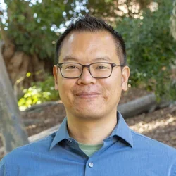
Photo of Jimmy Hu
Photo of Jimmy Hu

Jimmy Hu, PhD 

Study project: “Identifying progenitor populations and differentiation steps in developing orofacial structures using single cell epigenomic approaches”

Understanding how organs are normally formed and maintained is an important first step towards successful stem cell-based strategies for regenerative medicine, which will revolutionize our approach to treating diseases and alleviate problems caused by aging and trauma. The Hu Laboratory uses the tooth and other craniofacial structures as model systems to study fundamental principles of tissue morphogenesis during development and stem cell-based tissue renewal in adults. Project 1) Employed single cell RNA sequencing on dissociated mandibular epithelium and mesenchyme from mouse embryos. From this dataset, we hope to identify a) new putative markers and potential regulators for known progenitor populations; and b) new subsets of cells that may play important roles during dental and craniofacial development. Promising bioinformatic findings will be followed up with functional validation experiments. Project 2) We recently conducted spatial gene expression analysis of the embryonic mandible using the 10x Visium platform. Students will be delving into the dataset to unpack differential gene expression across the entire developing mandible. In addition, the same method was used on Msx1 null embryos and this will be a first step towards understanding how Msx1 regulates mandibular development. Msx1 mutation underlies several human craniofacial birth defects, including cleft lips and palates. Project 3) Understanding the regulation of adult stem cells in the mouse incisor, which is a highly trackable and accessible system for various genetic and imaging techniques. We have recently made a connection between cell shapes, tissue mechanics, and heterochromatin formation. Therefore, we are excited to conduct genomic studies, including single cell ATACseq, in order to test our hypothesis and to better understand how heterochromatin is regulated by tissue architecture and mechanical forces in adult stem cells.

Read More
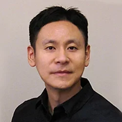
Photo of Dr. Yong Kim

Yong Kim, PhD

Study project: “Deep learning discovery of cancer-specific salivary cfDNA” 

Tremendous efforts have been devoted for the development of cancer-specific biomarkers based on 'omics' signatures. Salivary exRNA biomarkers including miRNAs hold promise for detecting various local and systemic diseases such as oral cancer, Sjögren syndrome, pancreatic cancer and lung cancer. Liquid biopsy (LB) on circulating cell free DNA (cfDNA) with cancer-associated genomic mutations has become a great interest in cancer detection. However, their reliability and predictive power must be improved to become the standard of clinical care. This project aims to examine physical and genetic signatures in salivary cfDNAs associated with human gastric cancer (GC), and relate molecular signatures to detailed clinical parameters such as gender, age, symptoms, diet, infection status, habitual characteristics. We will leverage saliva samples prospectively collected and stored in our repository as a model to examine physiognomies of saliva cfDNAs associated with GC. The overarching goal is to develop a comprehensive non-invasive diagnostic cassette for early detection of GC. The computational biology component of this project is to evaluate multi-variate and deep learning (e.g. neural nets) algorithms for their predictive power in relating GC-specific cfDNA characteristics with clinical/demographic data.

Read More
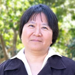
Photo of Dr. Hung Ton-That

Hung Ton-That, PhD

Study project: “Identifying and characterizing genetic variants of oral actinobacteria”

Oxidative protein folding is a general feature of Gram-positive actinobacteria, including the oral colonizers Actinomyces oris, Corynebacterium diphtheriae, and Corynebacterium matruchotii. In these organisms, a thiol- disulfide oxidoreductase named MdbA catalyzes post-translocational folding of exported proteins. Genetic disruption of mdbA causes cell arrest, altered cell morphology, antibiotic sensitivity, and attenuation of virulence, indicating that MdbA-mediated oxidative protein folding is an essential process in oral Actinobacteria. How this protein folding mechanism is linked to cell essentiality remains elusive. We propose to identify genetic suppressors that compensate the loss of mdbA. The genetic and biological nature of these suppressors will be revealed by a combination of whole genome sequencing and genetic and biochemical characterization. Computational analysis will first focus on identifying genetic variants from genome sequencing, and then to evaluate the potential functionality of these variants using network models.

Read More
Photo of Dr. Min Lee

Min Lee, PhD

Study project: “Enhanced gene therapy with targeted delivery systems”

Adeno-associated virus is one of the leading delivery vector for gene therapy in current clinical development due to its non-pathogenic potential and good safety profile in clinical trials. However, significant drawbacks exist after systemic delivery, including off-target gene transfer (e.g. liver sequestration) and low transduction of target tissue. Thus, this study will further enhance the efficacy and safety of viral vectored gene transfer by introducing therapeutic viruses into cell-derived exosomes modified for tissue targeting. The Lee lab will modify exosomes to increase targeting ability of therapeutic agents by employing genetic engineering of donor cells during exosome production or chemical conjugation of targeting ligands to exosome surface. In addition, they will perform non-viral mediated gene transfer by using non-phospholipid liposome nanocarriers (sterosomes) with high sterol content (50-70 mol%), leading to superior gene transfection efficiency compared to conventional phospholipid liposomes. They will also introduce alendronate onto the sterosomes surface for potential bone tissue targeting using its affinity of hydroxyapatite. This technique will be a useful tool for surface functionalization of delivery vehicles with various targeting moieties as well as imaging agents, providing a versatile vector platform for more efficient and safe gene therapy and drug delivery applications.

Read More
Photo of Dr. Yi-Ling Lin

Yi-Ling Lin, DDS, DMSc

Study project: “Identification of exRNA biomarkers for ossifying fibroma and cemento-osseous dysplasia”

Ossifying fibroma and cemento-osseous dysplasia are benign fibrous-osseous lesions (BFOLs) that normally occur in the craniofacial skeleton. In both lesions, normal bone is replaced by a cellular fibrous tissue containing various forms of ossification. Due to their overlapping histological features, the distinction cannot be made solely by histology without clinical and radiographic correlation. This poses a diagnostic challenge for the detection of ossifying fibroma at early stages. The distinction of these two lesions is important since they require very different treatment. Ossifying fibroma is a tumor that needs to be surgically resected, while cemento-osseous dysplasia is a developmental anomaly, which requires no active treatment. Therefore, the Lin lab plans to use a functional genomic approach to identify biomarkers that will enable ossifying fibroma to be distinguished from cemento-osseous dysplasia. Samples from their oral pathology archives will be used as the source material. The RNAs from the block sections will be extracted and processed for RNA sequencing analysis, which provides the platform to establish a global gene expression profile. The future comprehensive bioinformatic analysis will identify the genes that are significantly differentially expressed between the two lesions. These biomarkers may potentially have great values for the early diagnosis of ossifying fibroma and reducing bone destruction brought by the disease and surgery. In addition, the functional analysis of RNA sequencing data can also reveal aberrantly affected cellular pathways in ossifying fibroma and cemento-osseous dysplasia, potentially leading to new therapeutic strategies.

Read More
Photo of Dr. Renate Lux

Renate Lux, PhD

Study project: “Biofilms in oral health and disease”

The Lux laboratory is focused on the understanding of oral microbial communities and their role in oral health and disease. As part of this overall research interest, they have been studying the oral microbiome composition of biofilms associated with the gingival crevice in periodontal disease with a focus on how treatment effects the microbiome. Ongoing and future research projects related to this question include longitudinal studies of the microbiome before and after treatment, which allow the identification of molecular signatures with predictive power for risk of disease recurrence, the effect of type 2 diabetes and smoking as risk factors as well as the influence of adjunctive laser treatment on microbiome composition. In addition to the subgingival microbiome, the Lux lab is also studying microbiomes associated with artificial surfaces in the oral cavity including dentures, orthodontic appliances, etc. This includes longitudinal studies of the tooth-, gingiva and saliva-associated microbiomes in patients undergoing orthodontic treatment with either clear aligners or conventional brackets. This study evaluates and compares these microbiomes before treatment and at 1, 3, 6 and 12 months after beginning of the treatment and then follows the microbiome for six months after the patient is debonded. In a separate study, the lab investigates in detail the initial microbiome changes of the tooth- and gingival crevice-associated microbial communities on a weekly basis during the first month. In addition to microbiome, the Lux laboratory has started to examine the genomes of key oral microbial species such as Fusobacterium nucleatum isolated from healthy and diseased gingival site for differences in genomic content. The goal is to identify genomic content and signatures that are correlated with health- and disease association.

Read More
Photo of Dr. Alireza Moshaverinia

Alireza Moshaverinia DDS, MS, PhD, FACP

Study project: “Hydrogel biomaterial to direct the fate of the encapsulated MSCs in bone regeneration”

Mesenchymal stem cells (MSCs) are promising alternative treatment for bone regeneration. MSCs derived from orofacial tissues (e.g. gingival mesenchymal stem cells (GMSCs) are attractive postnatal stem cells with self- renewal, multilineage differentiation capacities, and comparable osteogenic properties to bone marrow MSCs (BMMSCs). GMSCs are readily accessible in the oral cavity and can be easily found in the discarded tissue samples. Studies have shown that proinflammatory T-cells and cytokines inhibit MSC-mediated bone tissue regeneration limiting their clinical application. It is well known that biomaterials are used to direct the fate of MSCs. However, controlling the fate of the transplanted MSCs is still a major challenge. The Moshaverinia lab is interested in understanding the factors influencing the fate of encapsulating MSCs. The elasticity of the hydrogel biomaterial is a crucial factor influencing MSC differentiation, but its role in the MSC-host immune system is fairly unknown. To develop effective MSC-mediated therapies it is crucial to have a clear understanding of how the encapsulating biomaterial elasticity affect the MSCs-host immune system interplay and MSCs immunoregulatory properties. Therefore, they aim to understand the role of elasticity of the hydrogel biomaterial to direct the fate of the encapsulated MSCs through regulation of dental MSCs-immune cells crosstalk; and to evaluate the effects of elasticity of the hydrogel biomaterial in MSC immunoregulatory properties. 

Read More
Photo of Dr. Ichiro Nishimura

Ichiro Nishimura DDS, PhD, DMSc, DMD

Study project: “Science of the Face”

This project seeks to enhance the student’s understanding of the challenge of planning, conducting and interpreting the scientific research in the field of biotechnology and genomics. The Nishimura lab will use the Face to investigate and try to answer the following questions: How is the face developed?; What are the facial abnormalities?, Does the face contribute to mutual understanding or more to misunderstanding?; What is the function of the face?; etc. It is well established that facial abnormalities are most frequent phenotypes of inherent diseases. Selected clinical cases highlighting the unique functions of the face will be investigated. Overall the genotype, phenotype and stereotype using the Face will be explored in detail.

Read More
Photo of Dr. Sotirios Tetradis

Sotirios Tetradis, DDS, PhD

Study project: “Genetic determinants of medication-related osteonecrosis of the jaws (MRONJ)”

Medication-related osteonecrosis of the jaws (MRONJ) is a significant side effect of antiresorptive medications, such as bisphosphonates (BPs) and denosumab (dmab) that can have devastating effects on patients’ well-being. BPs and dmab are used to inhibit osteoclastic function in conditions such as bone malignancy or osteoporosis. Despite described in the literature since 2003, MRONJ etiology and pathophysiology remain largely unknown. MRONJ pathogenesis appears multifactorial and affects only a small subset of patients. However, the specific characteristics of this susceptible subgroup are still unknown. Identifying patients at risk of developing MRONJ would allow modification of treatment protocols and application of preventive measures. As mice share structural, functional and genetic traits with humans and can be raised in a tightly controlled environment, the Tetradis lab will utilize the inbred mouse strains and their well-established MRONJ model to begin unraveling murine MRONJ genetics, with an eye toward future translational studies on genetic and environmental regulators of the human disease. They will utilize the Hybrid Mouse Diversity Panel (HMDP) of inbred mouse strains to explore genetic determinants of increased MRONJ risk. In addition, they intend to test the association of these markers in patients with MRONJ. Ultimately, with eyes on a personalized patient approach, they aim to identify patients in high risk and to develop prognostic and targeted therapeutic interventions for MRONJ. 

Read More
Photo of Dr. Cun-Yu Wang

Cun-Yu Wang, DDS, PhD

Study project: “Epigenetic mechanisms that control orofacial bone aging and osteoporosis"

The Wang’s Lab has two projects related to genome sciences and medicine to be performed. The first project is to study the epigenetic and molecular mechanisms that control orofacial bone aging and osteoporosis in parallel with long bones and to explore the extent to which targeting epigenetic factor could prevent orofacial bone aging and promote dental, oral and craniofacial tissue regeneration. Mesenchymal stem/stromal cells (MSCs), derived from orofacial bone tissues (OMSCs), are excellent sources for craniofacial bone regeneration due to their closer embryonic origins to the injured sites. In this project, we will explore how epigenetic factors will regulate orofacial bone aging and self-renewal of OMSCs in parallel with bone marrow MSCs. New findings from our studies will have important implications in developing innovative therapeutic strategies for preventing orofacial bone aging and promoting craniofacial bone regeneration. The second project is to develop novel therapeutics for head and neck squamous cell carcinoma (HNSCC) progression and metastasis. Emerging evidence suggests that histone methylation plays a critical role in activation of gene transcription in HNSCC by regulating chromatin accessibility. Recently, immune checkpoint inhibitors targeting PD1/PD-L1 have achieved good success in several solid tumors including melanoma and HNSCC. Although anti-PD1 therapy has been approved for treating recurrent or metastatic HNSCC, the objective response rate is less than 20%, indicating that HNSCC cells might be intrinsically resistant to checkpoint blockades. In this project, we will explore whether targeting epigenetic factors could activate tumor-intrinsic immunity and help to overcome HNSCC resistance to PD-1 blockade therapy by recruiting and activating CD8+ T cells. The results from our studies might have important implications for harnessing chromatin and epigenetic regulators for cancer immunotherapy by aggravating replication stress to induce tumor cell-intrinsic immune responses.

Read More
Photo of Dr. David Wong

David Wong, DMD, DMSc

Study project: “Salivaomics, exRNA, cfDNA, and machine learning for non-invasive early detection of cancer”

Gastric cancer (GC) begins in the stomach and is the fourth most common cancer occurring worldwide with an estimate of around 990,000 new cases each year. Gastric cancer detected at the early stage is more treatable and has a higher survival rate (65%), compared to the later stage (<20%). However, the diagnosis of GC is often delayed due to lack of early symptoms. And, imaging tests (CT, PET, and X-ray) currently used for the diagnosis of GC are not suitable for early cancer detection. Therefore, there is an urgent need for predictive biomarkers that can be used as a credible screening tool for early detection of GC. Saliva is an emerging biofluid for biomarker development for noninvasive detection and screening of local and systemic diseases, this project proposes to (1) profile genomic characteristics of salivary extracellular RNAs (exRNAs) associated with gastric cancer (200 saliva samples: 100 GC & 100 Control) and (2) perform machine learning development of gastric cancer specific genes, clinical and demographic characteristics (stage of GC, age, gender, ethnicity, smoking, H. pylori and EBV infection status, etc.). Employing a deep learning process with sufficient power will allow the development of the best fit algorithm that takes all related parameters into considerations and finds comprehensive signatures most specifically associated with gastric cancer, especially its early stage. Focus will be put on demonstrating the potential and efficacy of automated feature selection. In the era of big data science, we will concentrate on the potential of machine learning to facilitate the insight of cancer stage classification with major goal to enable early detection of the disease. Accomplishing this task will substantially alter the treatment for people suffering from GC as it will highly reduce the number of unnecessary endoscopies.

Read More