Embracing and Integrating AI
ADEA Grant for New Calibration Tool Means Greater Accuracy, More Cases, and Less Time Teaching the Nuances of Oral Radiology
This content appears in the UCLA School of Dentistry's spring 2025 magazine. Click here for all magazine content.
Teaching dental students to evaluate dental X-rays is laborious ... for both educators and pupils.
Students honing their skills “require repeated viewing of a variety of patients and contexts,” notes Dr. Sanjay Mallya, chair of the UCLA School of Dentistry’s Section of Oral and Maxillofacial Radiology.
Moreover, consistency in calibration can vary even among faculty.
“There can be subjectivity in interpretation leading to intraoperative variability,” says Dr. Kumar C. Shah, professor of clinical dentistry and residency program director for advanced prosthodontics.
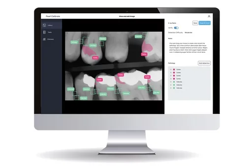
Artificial intelligence has the potential to remediate these issues dramatically. This year, the UCLA School of Dentistry received a grant from the American Dental Education Association Gies Foundation to incorporate and evaluate the first AI-powered clinical calibration tool. The Calibrate system from dental AI company Pearl combines a pathoses detection tool with an evaluation platform.
Before applying the system to D.D.S. student and resident education, Dr. Shah noted that a faculty team used it to look at consistency and calibration. “We had four different faculty looking at the same set of radiographs individually and with the assistance of AI, and we found that when the faculty used the AI as an aid, they were a lot more consistent among themselves,” he says.
They then used images calibrated by faculty to explore how the Calibrate system could help train students to interpret dental X-rays.
“You can teach anatomy and general principles of how caries might occur and how bone loss might appear. But student learning must be accomplished by repeatedly seeing radiographs from different patients with different anatomies in different contexts,” says Dr. Mallya. “It requires enabling students to refine their skills over time.”
Teaching these skills is faculty-intensive, and the investigators believe the program could alleviate some of that demand. “By enabling students to learn and calibrate themselves with less faculty oversight, we could address and alleviate bandwidth constraints as well,” says Dr. Shah.
The extensive built-in library of X-rays has undergone initial AI review, and these are validated by faculty for later use in a testing environment.
“Students see a limited patient pool over the course of their training, whereas the AI system can expose them to a wide range of images they can interact with in a one-on-one experience,” says Dr. Mallya.
The system can be used to have students identify common dental pathoses including decay, periapical infections in the jaw, and bone loss from periodontal disease. The program can be used to have a student identify dental decay, specify whether the decay occurs only in the enamel or has penetrated the dentin, and describe the depth of the decay. Faculty can then see whether a student tends to under or overestimate the area of decay, and the students themselves receive immediate feedback.
Calibrate enables images to be presented in a real-world context. It can also limit the time students have to analyze a particular image, preparing them for the time and productivity demands they’re likely to face in practice.
The ADEA-funded study involves recruiting dental students and examining the impact of using AI-based caries detection to facilitate student radiographic diagnostic skills. The next step of the grant involves piloting the program with entering third-year students and tracking student performance in their oral radiology rotation over the course of the year. It is expected to help faculty better identify student challenges and weaknesses and tailor lessons accordingly.
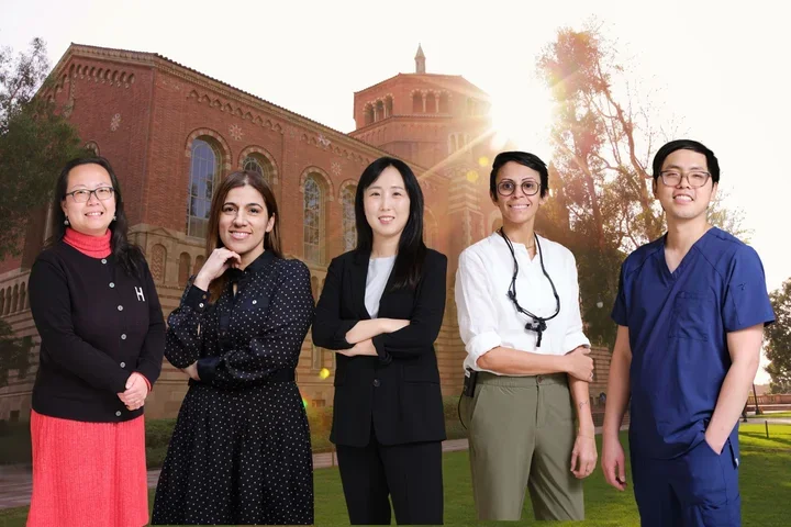
Nathan Fok, D.D.S. ’25, O.M.R. ’27, is in an advantageous position to fully explore Calibrate’s capabilities. Having already conducted predoctoral research on AI applications in detecting caries, he will now leverage the technology – and mentor UCLA dental students – while enrolled in the School’s two-year Oral and Maxillofacial Radiology residency program.
"Dental students starting out in the clinic may struggle to confidently detect cavities on radiographs on their own,” Fok said. “Calibrate can serve as a valuable training tool, reinforcing their interpretations, building diagnostic confidence, and helping them calibrate their eye to real clinical findings.”
Dr. Mallya notes that Calibrate has the potential to perform other types of assessments, such as faculty calibration across the School or across institutions. He envisions the School of Dentistry’s instructional models being potentially shared and adapted by other universities.